41 muscle fiber model with labels
Pin on Strength Training Benefits - Pinterest Muscle Fiber Model: Motor Neuron, Myeline Sheath, Node of Ranvier, Synaptic Terminal, Synaptic Cleft, Endomysium, Sarcolemma, Nuclei, Mitochondria, T-tubules, Sarcoplasmic Reticulum, Myofibrils Find this Pin and more on Strength Training Benefits by Great Bones. More like this A sarcomere is the functional unit of striated muscle. Sarcopenia: Aging-Related Loss of Muscle Mass and Function Jan 01, 2019 · A. Methodological Problems in the Study of Aging Skeletal Muscle. The study of sarcopenia in humans is complicated by the long duration of the aging process, large variability among individuals, and multiple factors affecting muscle that are not primarily related to aging per se. Studies of aging can be conducted using either a cross-sectional or longitudinal design, but neither are free from ...
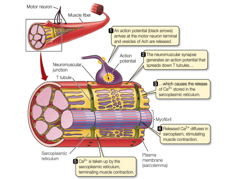
Muscle Fiber. 1. Myofibrils 2. Mitochondrium 3. Postsynaptic membrane 4 ... Muscle Fiber. 1. Myofibrils 2. Mitochondrium 3. Postsynaptic membrane 4. Synaptic gap with basal lamina 5. Presynaptic membrane 6. Presynaptic vesicle 7. Schwann cell 8. Nucleus 9. Actin filament 10. Sarcomere 11. Myosin filament 12. Myelin sheath 13. Neurofibers 14. Cell membrane (sarcolemma) 15. Transverse membrane tube 16. Triad 17.
Muscle fiber model with labels
anatomy labeled muscle fiber Muscle Cells - Types Of Cells In The Body jatypesofcells.weebly.com. muscle cell diagram cells parts. Sarcolemma lookfordiagnosis.com. sarcolemma improve help muscle fiber. Anatomy muscles posterior human chart muscle medical lithograph frame 1930s drawing structure revisit later favorites sold. Muscle cells. Skeletal Muscle Fiber Labeling - Printable About this Worksheet. This is a free printable worksheet in PDF format and holds a printable version of the quiz Skeletal Muscle Fiber Labeling.By printing out this quiz and taking it with pen and paper creates for a good variation to only playing it online. Muscles Labeling - The Biology Corner The activity linked below is a drag and drop activity for students to practice labeling the muscles, there are 6 slides showing images of muscles and fibers and the connective tissue surrounding the fibers (endomysium, perimysium, epimysium). Google Slides Key (TpT) Prev Article Next Article
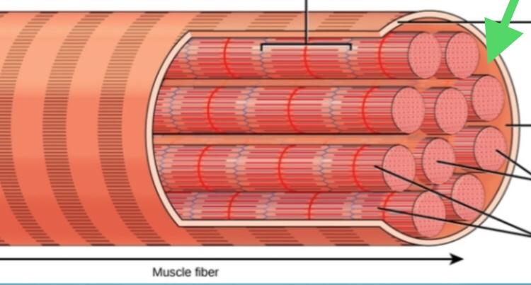
Muscle fiber model with labels. Skeletal Muscle Fiber Structure and Function - Open Textbooks for Hong Kong Each skeletal muscle fiber is a skeletal muscle cell. Within each muscle fiber are myofibrils, long cylindrical structures that lie parallel to the muscle fiber.Myofibrils run the entire length of the muscle fiber. They attach to the plasma membrane, called the sarcolemma, at their ends, so that as myofibrils shorten, the entire muscle cell contracts (Figure 16.18). Skeletal Muscle Fiber Model - Myofibrils - YouTube This video was produced to help students of human anatomy at Modesto Junior College study our anatomical models. muscle anatomy not labeled Histology respiratory trachea cartilage labeled perichondrium epithelium hyaline system tissue slides anatomy lamina propria embryology edu label tract labels human. Anatomy 99worksheets. Muscle junction neuromuscular fiber anatomy muscular system Skeletal Muscle Fiber Location and Composition - GetBodySmart A review of skeletal muscle fiber (cell) location, structure, anatomy, and function using interactive animations, models, and labeled diagrams. Start learning now!
Sarcomere Model | Muscle Fiber Model | Skeletal Muscle | MICROanatomy ... This micro-anatomy model magnifies the anatomy of the human muscle fiber approximately 10,000 times. This muscle model illustrates a section of a skeletal muscle fiber and its neuromuscular end plate. The muscle fiber is the basic element of the diagonally striped skeletal muscle. You've never seen a muscle fiber in this way! Muscular System Anatomy and Physiology - Nurseslabs Option C: A muscle is composed of numerous visible bundles called muscle fasciculi. A fasciculus is composed of several muscle cells or muscle fibers. Each muscle fiber is a single cylindrical cell that contains several nuclei located at the periphery of the muscle fiber. The cytoplasm of the muscle fiber contains numerous myofibrils. General Anatomy of Skeletal Muscle Fibers | GetBodySmart Skeletal Muscle Fiber Location and Arrangement. are located inside muscles, where they are organized into bundles called […] Internal Anatomy of Skeletal Muscle Fibers. An interactive quiz about the internal anatomy of skeletal muscle fibers, featuring illustrations-based multiple choice questions. Satellite Cells and the Muscle Stem Cell Niche - PMC In adults, the percentage of satellite cells in soleus muscle is generally two- to fourfold higher than that in tibialis anterior muscle or EDL muscle (190, 466, 500). Within the same muscle, the number of satellite cells found on slow muscle myofibers (type I) is generally higher than those on fast myofibers (type IIa and type IIb) ( 190 , 324 ...
Muscle Models | Muscle Figures | Musculature Models - 3B Scientific 3B MICROanatomy™ Human Muscle Fiber Model, 10,000 times magnified - 3B Smart Anatomy $ 339.00 Item: 1000213 [B60] This micro-anatomy model magnifies the anatomy of the human muscle fiber approximately 10,000 times. This muscle model illustrates a section of a skeletal muscle fiber and its neuromuscular end plate. 9.2A: Skeletal Muscle Fibers - Medicine LibreTexts Skeletal Muscle Fiber Structure. Myocytes, sometimes called muscle fibers, form the bulk of muscle tissue. They are bound together by perimysium, a sheath of connective tissue, into bundles called fascicles, which are in turn bundled together to form muscle tissue. Myocytes contain numerous specialized cellular structures which facilitate their ... Muscle Fiber Model #1 - Ohio University - Anatomy & Physiology Muscle Fiber Model #1 - Ohio University - Anatomy & Physiology Amazon.com: Fibercon (Caplets) Fiber Therapy for Regularity ... GENTLE EFFECTIVE RELIEF: Just as effective as fiber powders: no mixing, no mess. FiberCon does not ferment, so it will not cause gas or bloating. Produces bowel movement in 12-72 hours. NO CHEMICAL STIMULANTS: Acts like natural fiber to help control and maintain normal bowel function.
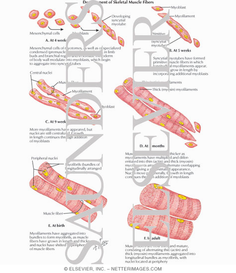
Development of Skeletal Muscle Fibers
Muscle Fiber Model Labeled Anatomy Muscle Fiber Model Labeled Anatomy Cardiac muscle fiber model. Sarcomere muscle skeletal bands band anatomy biology structure filament definition structural vs microscope associated muscular physiology isotropy cells below proteins.

Skeletal muscle fiber - Printable
Muscle Model - an overview | ScienceDirect Topics The muscle models have been used in the exoskeleton control schemes. Unlike the dynamic model, the muscle model predicts the muscle forces deployed by the muscles of the human limb joint as a function of muscle neural activities and the joint kinematics (Anam and Al-Jumaily, 2012). The input is the EMG signals and the output is force estimation.
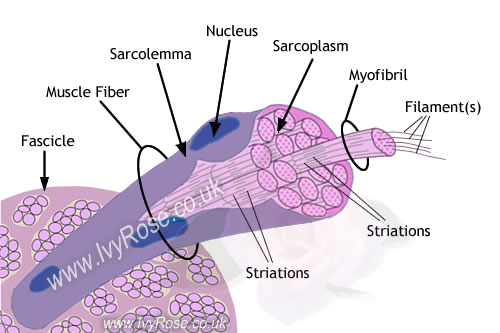
Muscle Fibre : Anatomy of Muscle Structure
Anabolic steroid - Wikipedia Most steroid users are not athletes. In the United States, between 1 million and 3 million people (1% of the population) are thought to have used AAS. Studies in the United States have shown that AAS users tend to be mostly middle-class men with a median age of about 25 who are noncompetitive bodybuilders and non-athletes and use the drugs for cosmetic purposes. "
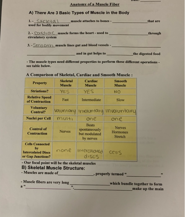
Solved: Anatomy Of A Muscle Fiber A) There Are 3 Basic Typ... | Chegg.com
Amazon.com: Muscle Milk Genuine Protein Shake, Chocolate, 11 ... Contains twelve (12) 11 fl oz Cartons of Muscle Milk Genuine Protein Shakes. Packaging may vary. HELPS SATISFY HUNGER AND BUILD MUSCLE – Muscle Milk Genuine is an energizing protein shake that can be consumed as an on-the-go breakfast or anytime snack or to support post-workout recovery and muscle growth.
Notes Muscles
Muscle Fiber Labeling Quiz - PurposeGames.com About this Quiz This is an online quiz called Muscle Fiber Labeling Quiz There is a printable worksheet available for download here so you can take the quiz with pen and paper. Your Skills & Rank Total Points 0 Get started! Today's Rank -- 0 Today 's Points One of us! Game Points 17 You need to get 100% to score the 17 points available
Ch. 9 Muscles and Muscle Tissue Flashcards | Easy Notecards
A 3D culture model of innervated human skeletal muscle enables studies ... 3D culture enhances skeletal muscle fiber maturation over 2D culture. (A) Representative confocal images of muscle fibers established in 2D (top row) and 3D conditions and immunostained for sarcomeric α-actinin (SAA; red), α-bungarotoxin (BTX; green), and Hoechst 33342 (blue) after 1, 2, and 3 weeks of culture.Scale bar, 50 μm. White arrowheads indicate broken fibers.

bone model labeled | Bone Model with labels : | Anatomy and physiology, Anatomy study, Anatomy
Skeletal Muscle Labeling MUSCULAR SYSTEM ANATOMY:Muscle Fiber With Neuromuscular Junction Model muscle junction neuromuscular fiber anatomy muscular system HLS [ Muscle Tissue, Skeletal Muscle, Ensheathment, Transverse muscle skeletal fascicles labeled histology tissue A Series Of Three Bones Showing The Basic Bone Markings. | Basic

Replika, Taman, Meubel, Jualan, Franchise, Pembantu, Pegawai, Mahasiswi, Guru, Gordyn, Jok ...
skeletal muscle anatomy labeled : Skeletal muscle fiber model, Skeletal muscle anatomy labeling part 1 - YouTube and also Neurolemmocyte On Skeletal Muscle Model - Human Anatomy - GUWS Medical. MRI Musculo-Skeletal Section: MRI Anatomy Of The Shoulder (sagittal View).

Human skeletal muscle fiber section - Stock Image - C005/1075 - Science Photo Library
Solved Exercise 2: Cell Structure From the information - Chegg Exercise 2: Cell Structure From the information provided in the preceding discussion, label the parts of the muscle fiber in Figure 11.4 2. 3. 6. (dark band) _(light band) 7. Figure 11.4: Model of muscle fiber.

32 Label The Structures Of A Skeletal Muscle Fiber - Label Design Ideas 2020
Muscle Fiber Model (Altay) Flashcards | Quizlet Muscle fiber model identifications Terms in this set (21) sarcolemma Identify the membrane. endomysium Identify the tissue layer. myofibril Identify the structure. thick myofilament Identify the structure. thin myofilament Identify the structure. neuromuscular junction Identify the connection. axon Identify the structure. axon terminals
Print Bio 201 Muscular System flashcards | Easy Notecards
PDF Anatomy & Physiology - TMCC Subcutis (Hypodermis) 1. External Horny Layer (Stratum corneum) 1a. Clear Layer (Stratum lucidum) -(KS 3 only) 2. Internal Hornless Germinative Zone (Stratum germinativum) 2a. Granular Layer (Stratum granulosum) 2b. Prickle-cell Layer (Stratum spinosum) 2c. Cylindrical Layer (Stratum basale) 3. Papillae 4.

Muscle fiber model labeled | A&P.2.Skin.Bone.Muscle | Pinterest | Muscle, Models and Smooth
Solved Lab 9: Muscle Tissue and axial muscle Exercise 1. | Chegg.com Identify and list a function of each labeled item 1-8) on the models Below. These are the same model at different angles. These are models of ONE muscle fiber or one skeletal muscle cell. Use the following terms to help you label: Endomysium, sarcolemma, myofibril, sarcoplasmic reticulum, myofilaments, motor neuron, T-tubule, nucleus.
Print Chapter 10 flashcards | Easy Notecards
SAC A&P Model Key - Muscular System Muscular System. M1 - Muscled Arm. M2 - Muscle Leg. M3 - Female Muscle Figure. M4 - Microanatomy Muscle Fiber. M5 - Muscle Figure.

Skeletal Muscle Fiber Organization
Muscle Cells (Myocyte) - Physiopedia In the anterior thigh, a muscle fiber may be a meter long. In contrast, muscle fibers making up the stapedius, a small muscle of the inner ear, are only a few millimeters in length. The Sarcoplasm Rich with glycogen (a form of stored energy) and myoglobin (a molecule that can store some O2), both required for energy generation

What are muscle fibers made of — a smaller muscle, such as those
Muscle Milk Genuine Protein Powder, Chocolate, 1.93 Pound, 12 ... Arrives by Fri, Jul 8 Buy Muscle Milk Genuine Protein Powder, Chocolate, 1.93 Pound, 12 Servings, 32g Protein, 2g Sugar, Calcium, Vitamins A, C & D, NSF Certified for Sport, Energizing, Workout Recovery, Packaging May Vary at Walmart.com

Diagram Of A Typical Muscle Fibre Gallery Muscle Fiber Image Unlabeled Human Anatomy Diagram Gif ...
Altay Size: 110x19x14,5 Weight: approx. 5100 g. Full Description. Muscular Body - cod:6000.58. This 1/4 life-size model is a useful tool to study human superficial musculature. Significant structures are numbered and referenced on the accompanying k-card. Size: 25x18x40 cm Weight: 765 g. Full Description. Skeletal Muscle Fiber - cod:6000.32. This ...
Post a Comment for "41 muscle fiber model with labels"