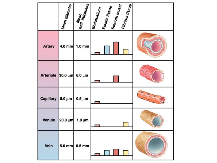
39 structure of the heart with labels
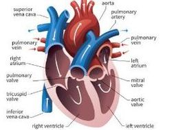
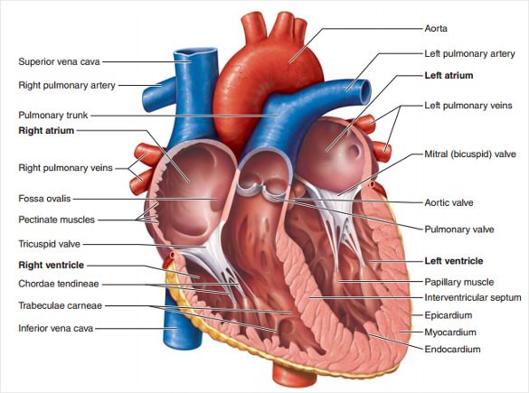
Human Heart - Diagram and Anatomy of the Heart - Innerbody The heart is a muscular organ about the size of a closed fist that functions as the body's circulatory pump. It takes in deoxygenated blood through the veins and delivers it to the lungs for oxygenation before pumping it into the various arteries (which provide oxygen and nutrients to body tissues by transporting the blood throughout the body). Heart Diagram with Labels and Detailed Explanation There are four chambers of the heart. The upper two chambers are the auricles and the lower two are called ventricles. There are four main valves of the human heart- aortic valve, mitral valve, pulmonary valve and tricuspid valve. They help prevent backflow of the blood.
Heart Labels - Printable or Custom Printed Stickers | Avery.com Use our free specialty shape label templates to easily personalize your heart labels online. Customize one of our free designs or upload your own graphics and then choose the printing option that works best for you. Order your blank heart labels or custom printed heart labels and stickers online and get free shipping on orders of $50 more.

Structure of the heart with labels
The structure of the heart - Structure and function of the … 14.04.2022 · It is located in the middle of the chest and slightly towards the left. The heart is a large muscular pump and is divided into two halves - … Heart Anatomy: Labeled Diagram, Structures, Function, and Blood Flow Chambers of the Heart Let's begin with the chambers of the heart. There are 4 chambers, labeled 1-4 on the diagram below. To help simplify things, we can convert the heart into a square. We will then divide that square into 4 different boxes which will represent the 4 chambers of the heart. Diagram of Human Heart and Blood Circulation in It 13.07.2022 · A heart diagram labeled will provide plenty of information about the structure of your heart, including the wall of your heart. The wall of the heart has three different layers, such as the Myocardium, the Epicardium, and the Endocardium. …
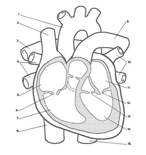
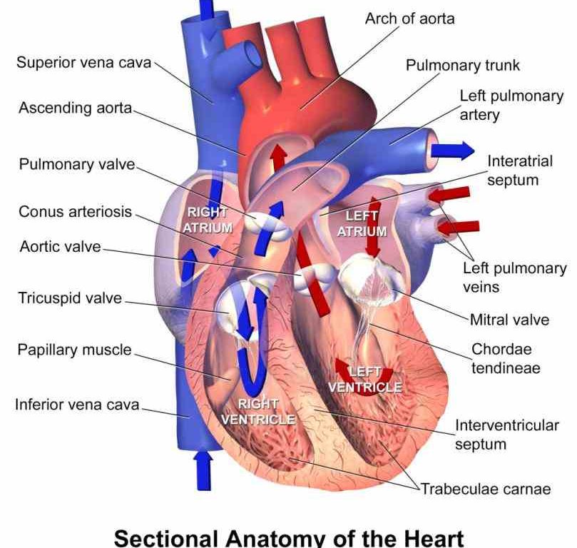
Structure of the heart with labels. Label the heart — Science Learning Hub Label the heart Interactive Add to collection In this interactive, you can label parts of the human heart. Drag and drop the text labels onto the boxes next to the diagram. Selecting or hovering over a box will highlight each area in the diagram. Right ventricle Right atrium Left atrium Pulmonary artery Left ventricle Pulmonary vein Semilunar valve Heart Anatomy Labeling Game - PurposeGames.com This is an online quiz called Heart Anatomy Labeling Game There is a printable worksheet available for download here so you can take the quiz with pen and paper. Your Skills & Rank Total Points 0 Get started! Today's Rank -- 0 Today 's Points One of us! Game Points 19 You need to get 100% to score the 19 points available Actions Heart Anatomy: Heart Dissection - University of Washington The picture below shows an anterior view of the heart with the pericardium removed. The letters indicated in the text refer to the labels on the picture. The anterior surface of the heart is characterized by the presence of the large arteries leaving the base of the heart, the pulmonary trunk (H) and the aorta (G). The pulmonary trunk is the ... Ch. 19 Circulatory System- heart Flashcards | Quizlet Place the labels in order denoting the flow of blood through the pulmonary circuit beginning with the right atrium and ending in the left atrioventricular valve. The first and last structures are given. Right atrium 1. tricuspid valve 2. right ventricle 3. pulmonary valve 4. pulmonary trunk 5. pulmonary artery 6. lungs 7. pulmonary vein
The Anatomy of the Heart, Its Structures, and Functions 05.04.2020 · The heart is situated within the chest cavity and surrounded by a fluid-filled sac called the pericardium. This amazing muscle produces electrical impulses that cause the heart to contract, pumping blood throughout the body. … Heart Diagram with Labels and Detailed Explanation 05.04.2022 · The heart is located under the ribcage, between the lungs and above the diaphragm. It weighs about 10.5 ounces and is cone shaped in structure. It consists of the following parts: Heart Detailed Diagram. Heart - Chambers There are four chambers of the heart. The upper two chambers are the auricles and the lower two are called ventricles. Heart Structure | BioNinja Heart Structure · Two atria (singular = atrium) – smaller chambers near top of heart that collect blood from body and lungs · Two ventricles – larger chambers ... heart anatomy questions heart label anatomy quiz background following please purposegames. External gross anatomy of the heart: anterior view. Costosternal anatomy describes the muscles and joints of the chest.. Intraperitoneal and retroperitoneal organs • digestive • anatomyzone. Random Posts.
Heart Blood Flow | Simple Anatomy Diagram, Cardiac Circulation ... - EZmed Step 1 and 6 involve a blood vessel, which makes sense as this is how blood enters and exits that side of the heart. Steps 2-5 involve a chamber, valve, chamber, and valve. So if you remember this general pattern, it will help you recall the order in which blood flows through each side of the heart. Label the Heart Diagram | Quizlet Septum. ... Right Atrium. ... Semi-lunar Valves. ... Left Pulmonary Veins. ... Right Pulmonary Veins. heart | Structure, Function, Diagram, Anatomy, & Facts The heart consists of several layers of a tough muscular wall, the myocardium. A thin layer of tissue, the pericardium, covers the outside, and another layer, ... Heart: Anatomy and Function - Cleveland Clinic The parts of your heart are like the parts of a house. Your heart has: Walls. Chambers (rooms). Valves (doors). Blood vessels (plumbing). Electrical conduction system (electricity). Heart walls Your heart walls are the muscles that contract (squeeze) and relax to send blood throughout your body.
The Circulatory System
Diagrams, quizzes and worksheets of the heart | Kenhub Worksheet showing unlabelled heart diagrams. Using our unlabeled heart diagrams, you can challenge yourself to identify the individual parts of the heart as indicated by the arrows and fill-in-the-blank spaces. This exercise will help you to identify your weak spots, so you'll know which heart structures you need to spend more time studying ...

Circulatory System Diagram | New Health Advisor
How to Draw the Internal Structure of the Heart (with Pictures) To draw the internal structure of a human heart, follow the steps below. Part 1 Finding a Diagram 1 To find a good diagram, go to Google Images, and type in "The Internal Structure of the Human Heart". Find an image that displays the entire heart, and click on it to enlarge it. 2 Find a piece of paper and something to draw with.
Get Structure Of Heart Diagram Gcse Background | World of Images
The Anatomy of the Heart, Its Structures, and Functions The heart is the organ that helps supply blood and oxygen to all parts of the body. It is divided by a partition (or septum) into two halves. The halves are, in turn, divided into four chambers. The heart is situated within the chest cavity and surrounded by a fluid-filled sac called the pericardium. This amazing muscle produces electrical ...

The Human Egg Cell Explained For Egg Donors - Altrui Egg Donation Agency
Human Heart Diagram Labeled | Science Trends 02.01.2019 · List Of Heart Structures. Heart Chambers. Ventricles – The bottom two heart chambers. Atra – The upper two heart chambers. Wall Of The Heart. …
Heart Labeling (Internal)
A Labeled Diagram of the Human Heart You Really Need … The human heart resembles the shape of an upside-down pear, weighing between 7-15 ounces, and is little larger than the size of the fist. It is enclosed in a bag-like structure called the pericardium, and is located between the lungs, …

38 Label The Following Diagram Of The Heart - Labels 2021
Heart anatomy: Structure, valves, coronary vessels | Kenhub The heart is shaped as a quadrangular pyramid, and orientated as if the pyramid has fallen onto one of its sides so that its base faces the posterior thoracic wall, and its apex is pointed toward the anterior thoracic wall.
called myocardium science External Structure Of Human Heart Anatomy structure of human heart ...
Structure of the Heart | SEER Training The human heart is a four-chambered muscular organ, shaped and sized roughly like a man's closed fist with two-thirds of the mass to the left of midline. The heart is enclosed in a pericardial sac that is lined with the parietal layers of a serous membrane. The visceral layer of the serous membrane forms the epicardium. Layers of the Heart Wall

Label Heart Anatomy Diagram Printout - EnchantedLearning.com Oxygen-poor blood enters the right atrium of the heart (via veins called the inferior vena cava and the superior vena cava). The blood is then pumped into the right ventricle and then through the pulmonary artery to the lungs, where the blood is enriched with oxygen (and loses carbon dioxide).

Label Function Human Heart Diagram And Function - Aflam-Neeeak
Label the Heart - The Biology Corner Shows a picture of a heart with letters and blanks for practice with labeling the parts of the heart and tracing the flow of blood within the heart.
Know the structure of the heart - Labelled diagram
Heart Labeling Quiz: How Much You Know About Heart Labeling? Here is a Heart labeling quiz for you. The human heart is a vital organ for every human. The more healthy your heart is, the longer the chances you have of surviving, so you better take care of it. Take the following quiz to know how much you know about your heart. Questions and Answers 1. What is #1? 2. What is #2? 3. What is #3? 4. What is #4?
13+ Heart Diagram Templates – Sample, Example, Format Download | Free & Premium Templates
The structure of the heart - Structure and function of the heart ... The heart is a large muscular pump and is divided into two halves - the right-hand side and the left-hand side. The right-hand side of the heart is responsible for pumping deoxygenated blood to ...
The Anatomy and Physiology of Animals/Respiratory System Worksheet - WikiEducator
Structure of the Heart | The Franklin Institute The heart consists of four chambers: two atria on the top and two ventricles on the bottom. Looking at the Valentine's Day heart, the two rounded humps at the top are rounded like the top of a lower-case "a." The bottom is shaped like a "v." Feel it working What else is inside your heart?
How would you label the structures (both external and internal) of a dissected pig's heart? - Quora
Label Internal Anatomy of The Heart Diagram | Quizlet Start studying Label Internal Anatomy of The Heart. Learn vocabulary, terms, and more with flashcards, games, and other study tools.
Label the heart - Teaching resources
Structure of the Heart | SEER Training Structure of the Heart. The human heart is a four-chambered muscular organ, shaped and sized roughly like a man's closed fist with two-thirds of the mass to the left of midline. The heart is enclosed in a pericardial sac that is lined with …

Biology A&P Lab: Study guide for quiz on Feb.25th
Heart Diagram with Labels and Detailed Explanation - BYJUS It pumps blood from the heart to different parts of the body and back to the heart. The most common heart attack symptoms or warning signs are chest pain, breathlessness, nausea, sweating etc. The diagram of heart is beneficial for Class 10 and 12 and is frequently asked in the examinations.
Post a Comment for "39 structure of the heart with labels"