39 eye diagram with labels and functions
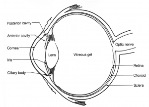
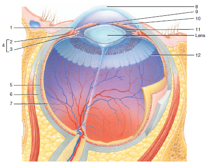
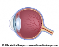
Eye Anatomy: 16 Parts of the Eye & Their Functions The following are parts of the human eyes and their functions: 1. Conjunctiva The conjunctiva is the membrane covering the sclera (white portion of your eye). The conjunctiva also covers the interior of your eyelids. Conjunctivitis, often known as pink eye, occurs when this thin membrane becomes inflamed or swollen. Eye Diagram With Labels and detailed description - BYJUS A brief description of the eye along with a well-labelled diagram is given below for reference. Well-Labelled Diagram of Eye The anterior chamber of the eye is the space between the cornea and the iris and is filled with a lubricating fluid, aqueous humour. The vascular layer of the eye, known as the choroid contains the connective tissue.
Labeled Eye Diagram | Science Trends What you want to interpret as a major part of the human eye is somewhat up to the individual, but in general there are seven parts of the human eye: the cornea, the pupil, the iris, the lens, the vitreous humor, the retina, and the sclera. Let's take a closer look at each of these components individually. The Cornea
Eye diagram with labels and functions
The Eye Diagram: What is it and why is it used? The eye diagram is used primarily to look at digital signals for the purpose of recognizing the effects of distortion and finding its source. To demonstrate using a Tektronix MDO3104 oscilloscope, we connect the AFG output on the back panel to an analog input channel on the front panel and press AFG so a sine wave displays. Then we press Acquire. Labeled Eye Diagram | Eye anatomy diagram, Eye anatomy ... - Pinterest accessory structures of the eye, extrinsic eye muscles, anatomy of the eyeball and microscopic anatomy of the retina. The skeletal system consists of bones and their associated connective tissues, including cartilage, tendons, and ligaments. It consists of dynamic, living tissues that are capable of growth, detect pain stimuli, adapt to stress ... Eye Anatomy Diagram - EnchantedLearning.com Retina - light-sensitive tissue that lines the back of the eye. It contains millions of photoreceptors (rods and cones) that convert light rays into electrical impulses that are relayed to the brain via the optic nerve. Rods - cells the in the retina that sense brightness (they are photoreceptors). Night vision involves mostly rods (not cones).
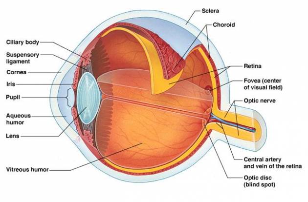
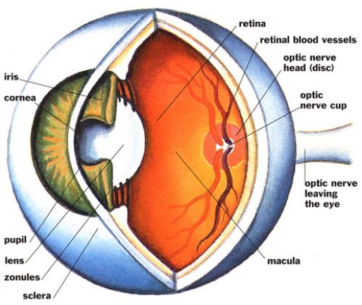
Eye diagram with labels and functions. Eye pattern - Wikipedia In telecommunication, an eye pattern, also known as an eye diagram, is an oscilloscope display in which a digital signal from a receiver is repetitively sampled and applied to the vertical input, while the data rate is used to trigger the horizontal sweep. It is so called because, for several types of coding, the pattern looks like a series of eyes between a pair of rails. BYJUS BYJUS Parts Of The Eye Labeled Diagram Model And Their Function The back layer or innermost parts of the eye are responsible for seeing and focusing. The back layer is made up of different parts: the retina, choroid, fovea, macula, and sclera. The retina is a thin tissue that covers the back of the eye. It sends images to the brain through the optic nerve. Label Parts of the Human Eye - University of Dayton Parts of the Eye Select the correct label for each part of the eye. The image is taken from above the left eye. Click on the Score button to see how you did. Incorrect answers will be marked in red.
PDF Parts of the Eye - National Eye Institute | National Eye Institute To understand eye problems, it helps to know the different parts that make up the eye and the functions of these parts. Here are descriptions of some of the main parts of the eye: ... Handout illustrating parts of the eye Keywords: parts of the eye, eye diagram, vitreous gel, iris, cornea, pupil, lens, optic nerve, macula, retina ... The Eyes (Human Anatomy): Diagram, Optic Nerve, Iris, Cornea ... - WebMD Articles On Eye Basics. Your eye is a slightly asymmetrical globe, about an inch in diameter. The front part (what you see in the mirror) includes: Iris: the colored part. Cornea: a clear dome ... Eye Anatomy | Definition, Structure & Functions - iBiologia Diagram of Human Eye with Labelling. Eye Anatomy Complete Physiology of Eye is described below in the given paragraph: The eye is rather like a living Camera. Each eye is a liquid-filled ball 2.5 cm in diameter. At the front of the eye is a clear, round window called the cornea. Behind the cornea is a "lens. PDF Eye Anatomy Handout - National Eye Institute of light entering the eye. Lens: The lens is a clear part of the eye behind the iris that helps to focus light, or an image, on the retina. Macula: The macula is the small, sensitive area of the retina that gives central vision. It is located in the center of the retina. Optic nerve: The optic nerve is the largest sensory nerve of the eye.
The Anatomy of Human Eye with Diagram | EdrawMax Online 1. The Anatomy of Human Eye. The most complex sensory organs of the human body are the eyes. Every part of the human body is responsible for a specific action, from the muscles and tissues to the nerves and the blood vessels. The human eye consists of many muscles and tissues that join to form an approximately spherical structure. Labelling the eye — Science Learning Hub Labelling the eye Add to collection The human eye contains structures that allow it to perceive light, movement and colour differences. In this activity, students use online or paper resources to identity and label the main parts of the human eye. By the end of this activity, students should be able to: identify the main parts of the human eye Eye Anatomy: Parts of the Eye and How We See Behind the anterior chamber is the eye's iris (the colored part of the eye) and the dark hole in the middle called the pupil. Muscles in the iris dilate (widen) or constrict (narrow) the pupil to control the amount of light reaching the back of the eye. Directly behind the pupil sits the lens. The lens focuses light toward the back of the eye. Human Eye Diagram, How The Eye Work -15 Amazing Facts of Eye First, light rays enter the eye through the cornea, the clear front "window" of the eye. The dome shaped cornea bends light to help the eye focus. From the cornea, the light passes through an opening called the pupil. The amount of light passing through is controlled by the iris, or the colored part of your eye.
NCERT Solutions for Class 8 Science Chapter 16 - Light - Arinjay Academy
Basic Eye Anatomy - Cataract Surgery Information The eyelashes also perform a protective function: they prevent dirt and other debris, as well as bacteria and viruses, from entering the eye, and are, therefore, very important in maintaining a healthy ocular surface. Sclera. The sclera is the white substance that makes up the wall of the eye. It is very dense and strong.

8 Best Images of Lens Diagram Worksheet - Microscope with Labeled Parts, Label Eye Parts ...
Labelling the eye — Science Learning Hub In this interactive, you can label parts of the human eye. Use your mouse or finger to hover over a box to highlight the part to be named. Drag and drop the text labels onto the boxes next to the eye diagram If you want to redo an answer, click on the box and the answer will go back to the top so you can move it to another box.

parts – Graph Diagram
Anatomy of the eye: Quizzes and diagrams - Kenhub One of our favorite ways to get to grips with all of the parts of the eye is by utilizing labeled diagrams. On a diagram of the eye, we can see all of the relevant structures together on one image. This helps us to understand how each one is situated and related to the other. Labeled diagram of the eye
Parts of the Eye and Their Functions | IYTmed.com
Eye anatomy: A closer look at the parts of the eye The iris of the eye functions like the diaphragm of a camera, controlling the amount of light reaching the back of the eye by automatically adjusting the size of the pupil (aperture). The eye's crystalline lens is located directly behind the pupil and further focuses light.
8 Best Images of Lens Diagram Worksheet - Microscope with Labeled Parts, Label Eye Parts ...
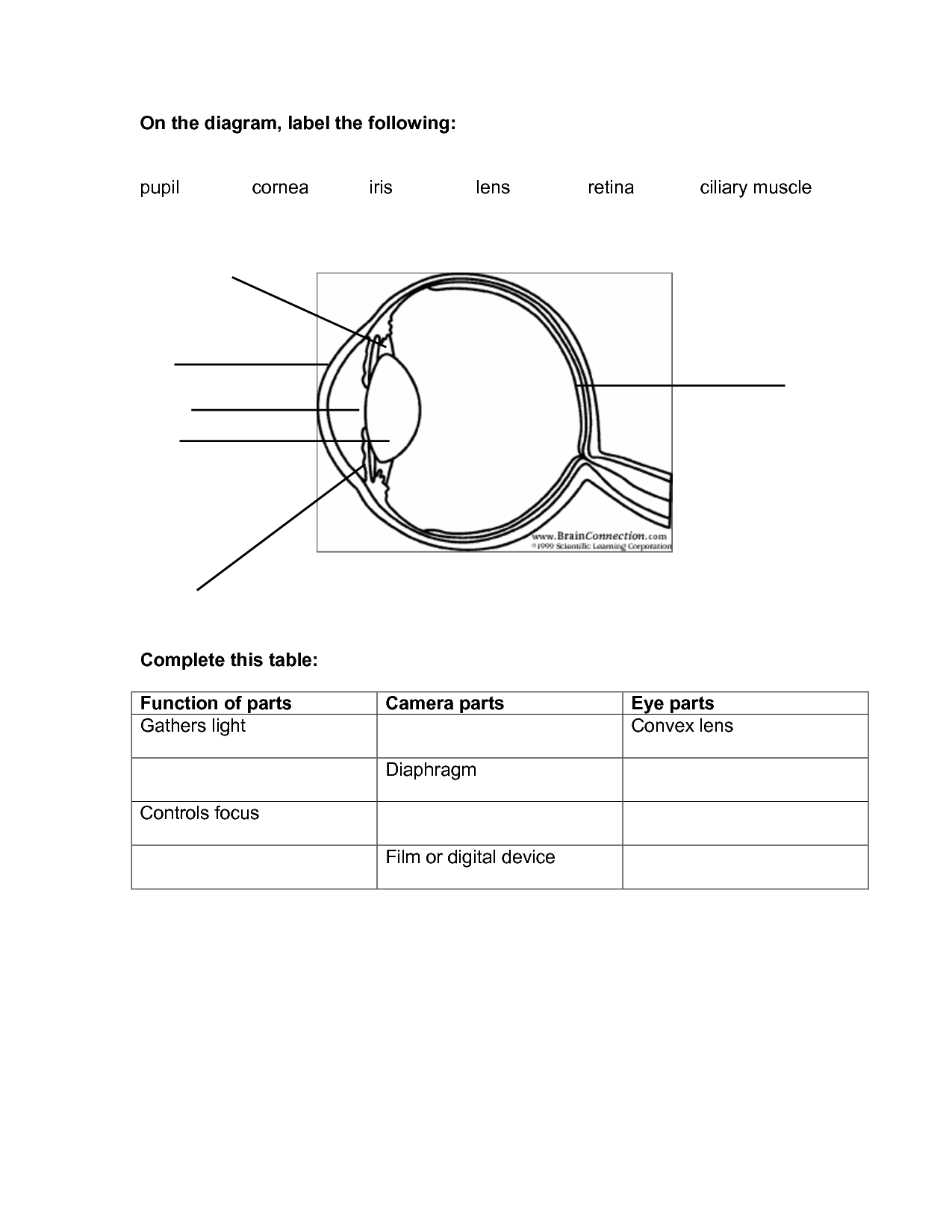
Draw a labeled diagram of human eye. Write the functions of Cornea ... Cornea of the eye is the first sight where convergence of light rays takes place. Iris is that part of the eye which controls the amount of light entering the eye through the pupil. Pupil is a type of small hole through which light enters the eye. The eye lens is a convex lens just behind the pupilwhich converges the light rays towards the ratina.

Pin on Drawings - Drapery & Perspective sight
Diagram of the Eye - Home - Lions Eye Institute In order for the eye to work at its best, all parts must work well collectively. To understand the eye and its functions, it's important to understand how the eye works, see below diagrams for both the external eye and the internal eye. The External Eye Instructions Click the parts of the eye to see a description for each.

A human labelled eye diagram!! A.M arts - YouTube
Structure And Function Of The Eye - Vision - MCAT Content The iris, which is conspicuous as the colored part of the eye, is a circular muscular ring lying between the lens and cornea that regulates the amount of light entering the eye. In conditions of high ambient light, the iris contracts, reducing the size of the pupil at its center. In conditions of low light, the iris relaxes and the pupil enlarges.

GK Questions and Answers on Human Eye
Structure of Human Eye (With Diagram) | Human Body The function of the eyebrows is to protect the anterior aspect of the eyeball from sweat, dust and other foreign bodies. 2. The Eyelids (Palpebrae) and Eyelashes: The eyelids are two movable folds situated above and below front of the eye. On their free edges, there are outgrowths of hairs— the eyelashes.

Human Body Pictures, Anatomy Diagrams, Free Science Images, Photos, Pics
Human Eye: Structure of Human Eye (With Diagram) | Biology The human eye is a very sensitive and delicate organ suspended in the eye socket which protects it from injuries. It essentially consists of CORNEA, LENS & RETINA besides many other parts such as Iris, Pupil and aqueous humour, vituous humour etc. Each one has got a specific function. A section of the eye is as shown in Fig. 2.2. ADVERTISEMENTS:
Free Blank Eye Diagram, Download Free Blank Eye Diagram png images, Free ClipArts on Clipart Library
Labelled Diagram of Human Eye, Explanation and Function - VEDANTU The basic functions of Rods and Cones are conscious light perception, color differentiation and depth perception. The human eye is capable of distinguishing between about 10 million colors, and it can also detect a single photo. The human eye is a part of the sensory nervous system. Labeled Diagram of Human Eye

Human Ear Diagram Blank - Diagram Media
Eye Anatomy Diagram - EnchantedLearning.com Retina - light-sensitive tissue that lines the back of the eye. It contains millions of photoreceptors (rods and cones) that convert light rays into electrical impulses that are relayed to the brain via the optic nerve. Rods - cells the in the retina that sense brightness (they are photoreceptors). Night vision involves mostly rods (not cones).
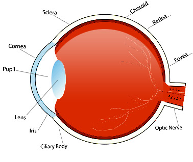
Diagram Of The Eye With Labels - ClipArt Best
Labeled Eye Diagram | Eye anatomy diagram, Eye anatomy ... - Pinterest accessory structures of the eye, extrinsic eye muscles, anatomy of the eyeball and microscopic anatomy of the retina. The skeletal system consists of bones and their associated connective tissues, including cartilage, tendons, and ligaments. It consists of dynamic, living tissues that are capable of growth, detect pain stimuli, adapt to stress ...

Glossary of Terms/Index - Screening for Schools
The Eye Diagram: What is it and why is it used? The eye diagram is used primarily to look at digital signals for the purpose of recognizing the effects of distortion and finding its source. To demonstrate using a Tektronix MDO3104 oscilloscope, we connect the AFG output on the back panel to an analog input channel on the front panel and press AFG so a sine wave displays. Then we press Acquire.

Diagram Of The Eye Not Labeled - Diagram Media
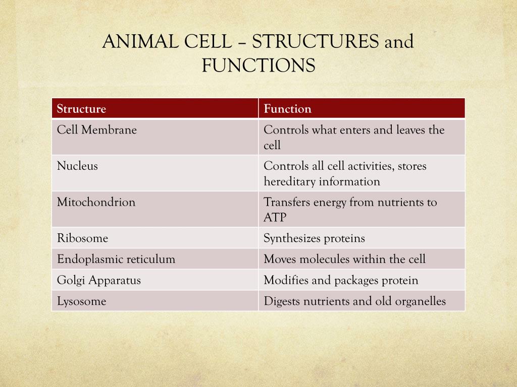
PPT - Cell Theory, Animal Cell Structure, Function, and Processes PowerPoint Presentation - ID ...
30 Label The Structure Of The Eye - Labels Database 2020

Eyes, Health and Eye anatomy on Pinterest
Simple Labeled Human Eye Diagram - Aflam-Neeeak
Post a Comment for "39 eye diagram with labels and functions"